Anatomy Diagram Rib Area / Rib Cage Organ Thoracic Cavity Internal Thoracic Artery Organs Heart Lung Anatomy Png Pngwing. Rib 1 is also flattened horizontally. Each pair is numbered based on their attachment to the sternum, a bony process at the front of the rib cage which serves as an anchor point. Several muscles that move the arms, head, and neck have their origins on the sternum. For more anatomy content please follow us and visit our website: The anatomy of the human ribs (costae) are one of the integral parts of the chest wall;
Anatomy diagram rib area : Its functions are to protect the thoracic organs from trauma and also form the bony attachment for various muscles. These pains may be stemming from the right or. Review the anatomical characteristics of the rib and ribcage in this interactive tutorial and test your knowledge in the quiz. Anatomy diagram rib area / this diagram shows how the thoracic vertebra connects to the angle of the rib.
Rib Cage Human Anatomy Organs from www.medicalook.com Each pair is numbered based on their attachment to the sternum, a bony process at the front of the rib cage which serves as an anchor point. Numbered ribs, sternum, cartilage parts and clavicular articulation. The sternum is located in the center of the anterior thoracic wall and is also known as the breastbone. The ribs are a set of twelve paired bones which form the protective 'cage' of the thorax. It is made up of 12 pairs of ribs. The bones of the pelvis and lower back work together to support the body's weight, anchor the abdominal and hip muscles, and protect the delicate vital organs of the vertebral and abdominopelvic cavities. Diagram of human body, liver rib cage, rib cage diagram labeled, rib cage diagram numbered, rib cage diaphragm, rib cage heart, rib cage organs anatomy, rib cage pain, stomach, diagram of human body, liver rib cage, rib cage diagram labeled, rib cage diagram numbered, rib cage diaphragm, rib cage. This article looks at female body parts and their functions, and it provides an interactive diagram.
The bones of the rib cage are the sternum, the 12 thoracic vertebrae and the 12 pairs of ribs.
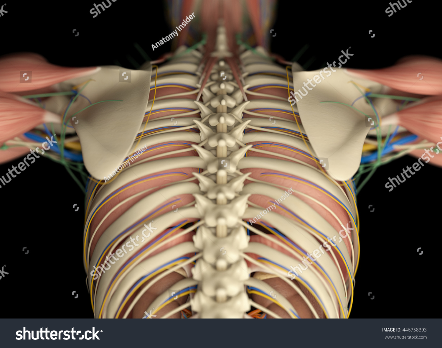
Rib cage, in vertebrate anatomy, basketlike skeletal structure that forms the chest, or thorax, and is made up of the ribs and their corresponding attachments to the sternum (breastbone) and the vertebral column.the rib cage surrounds the lungs and the heart, serving as an important means of bony protection for these vital organs.in total, the rib cage consists of the 12 thoracic vertebrae and. They make up the lateral part of our body, its anterior and posterior wall and they entirely build the lateral parts of the chest wall. Pain rib anatomy names human ribs cages injury rib cage art rib cage and pelvis sternum anatomy diagram. The vertebral column of the lower back includes the five lumbar vertebrae, the sacrum, and the coccyx. Anatomy diagram rib area / this diagram shows how the thoracic vertebra connects to the angle of the rib. The top edge of the manubrium has a depression called the suprasternal or jugular notch. The major muscles of the abdomen include the rectus. The bones of the pelvis and lower back work together to support the body's weight, anchor the abdominal and hip muscles, and protect the delicate vital organs of the vertebral and abdominopelvic cavities. Rib 2 is thinner and longer than rib 1, and has two articular facets on the head as normal. Anatomy of the rib cage diagram. In this image, you will find thoracic vertebrum, costochondral joint, costal cartilage, costal margin, costal arch, thoracic vertebrum, xiphoid process, xiphisternal joint, body, manubrial sternal joint, manubrium, the sternal notch in it. Vital organs such as heart and lungs are protected by the rib cage. The human rib cage is made up of 12 paired rib bones;
The sternum, commonly known as the breastbone, is a long, narrow flat bone that serves as the keystone of the rib cage and stabilizes the thoracic skeleton. The ribs are a set of twelve paired bones which form the protective 'cage' of the thorax. Bones of the pelvis and lower back. This bony framework plays an essential role in protecting the organs that lie in the thoracic region. Our latest youtube film is ready to run.
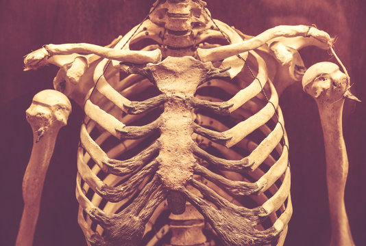
Human Anatomy Back Rib Cage Torso Stock Illustration 446758393 from image.shutterstock.com Diagram of human body, liver rib cage, rib cage diagram labeled, rib cage diagram numbered, rib cage diaphragm, rib cage heart, rib cage organs anatomy, rib cage pain, stomach, diagram of human body, liver rib cage, rib cage diagram labeled, rib cage diagram numbered, rib cage diaphragm, rib cage. The primary responsibilities of the ribcage involve protecting the thoracic visceral organs, enclosing the thoracic visceral organs, and is included. Of all 24 ribs, the first seven pairs are often labeled as 'true.'. It has a roughened area on its upper surface, from which the serratus anterior muscle originates. Its functions are to protect the thoracic organs from trauma and also form the bony attachment for various muscles. These muscles help the body bend at the waist. Female anatomy includes the external genitals, or the vulva, and the internal reproductive organs. It is made up of 12 pairs of ribs.
It has clear front side and back planes.
Structure of the ribcage and ribs. The heads of ribs 1, 10, 11, and 12 have a single facet for articulation with the bodies of the thoracic vertebrae. Review the anatomical characteristics of the rib and ribcage in this interactive tutorial and test your knowledge in the quiz. True ribs, false and floating. It has a roughened area on its upper surface, from which the serratus anterior muscle originates. The rib cage is joined to the thoracic vertebrae. Rib cage anatomy the rib cage, shaped in a mild cone shape and more flexible than most bone sets, is made up of varying elements such as the thoracic vertebra, 12 equally paired ribs, costal cartilage, and held together anteriorly by the sternum. Related posts of anatomy of ribs and its related area abdominal artery anatomy. Inspiration and expiration anatomical vector illustration diagram, educational medical scheme with lungs, diaphragm, rib cage and trachea. Rib 2 is thinner and longer than rib 1, and has two articular facets on the head as normal. The primary responsibilities of the ribcage involve protecting the thoracic visceral organs, enclosing the thoracic visceral organs, and is included. Anatomy of the rib cage diagram. The anatomy of the human ribs is made up of 24 ribs which are parted in 12 pairs (each on the left and right side of the chest wall), with the sternum, metasternum(the.
The bones of the rib cage are the sternum, the 12 thoracic vertebrae and the 12 pairs of ribs. It is also the center around which the superior 10 ribs directly or indirectly attached. Anatomy of the rib cage diagram. The major muscles of the abdomen include the rectus. The cartilage that forms at the end of each rib (costal cartilage) attaches either.
8 970 Best Human Rib Cage Images Stock Photos Vectors Adobe Stock from t3.ftcdn.net Of all 24 ribs, the first seven pairs are often labeled as 'true.'. This bony framework plays an essential role in protecting the organs that lie in the thoracic region. The sternum is located in the center of the anterior thoracic wall and is also known as the breastbone. Anatomy of the rib cage diagram. This article looks at female body parts and their functions, and it provides an interactive diagram. Rib 1 is also flattened horizontally. The rib cage is a bony structure found in the chest (thoracic cavity). The bones of the rib cage are the sternum, the 12 thoracic vertebrae and the 12 pairs of ribs.
The rib cage is joined to the thoracic vertebrae.
Abdominal artery anatomy 12 photos of the abdominal artery anatomy abdomen arteries anatomy, abdominal aortic anatomy, abdominal aortic aneurysm anatomy, abdominal aortic branches anatomy, abdominal vascular anatomy radiology, human anatomy, abdomen arteries anatomy, abdominal aortic anatomy, abdominal aortic. The primary responsibilities of the ribcage involve protecting the thoracic visceral organs, enclosing the thoracic visceral organs, and is included. The rib cage is the arrangement of ribs attached to the vertebral column and sternum in the. We are pleased to provide you with the picture named heart, lung, diaphragm and ribs location.we hope this picture heart, lung, diaphragm and ribs location can help you study and research. This bony framework plays an essential role in protecting the organs that lie in the thoracic region. Bones of the pelvis and lower back. See more ideas about anatomy, anatomy study, rib cage anatomy. The heads of ribs 1, 10, 11, and 12 have a single facet for articulation with the bodies of the thoracic vertebrae. These pains may be stemming from the right or. Our latest youtube film is ready to run. The sternum, commonly known as the breastbone, is a long, narrow flat bone that serves as the keystone of the rib cage and stabilizes the thoracic skeleton. Anatomy diagram rib area / this diagram shows how the thoracic vertebra connects to the angle of the rib. The ribs are a set of twelve paired bones which form the protective 'cage' of the thorax.
Share this post
0 Response to "Anatomy Diagram Rib Area / Rib Cage Organ Thoracic Cavity Internal Thoracic Artery Organs Heart Lung Anatomy Png Pngwing"

0 Response to "Anatomy Diagram Rib Area / Rib Cage Organ Thoracic Cavity Internal Thoracic Artery Organs Heart Lung Anatomy Png Pngwing"
Post a Comment